 TheIBAxolotl



Amino acids
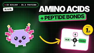
Every amino acid has the same core: a central carbon bonded to an amine group (NH₂), a carboxyl group (COOH), a hydrogen atom and a variable side chain called the R-group. The R-group is what makes each amino acid distinct. Proteins in living things use 20 amino acids coded for by the genetic code. Some are essential for humans: we cannot make them and must obtain them from food, so diet matters, for instance for vegans planning protein sources.
Peptide bonds
Two amino acids join by condensation between the carboxyl group of one and the amine group of the next. A peptide bond forms and one water molecule is released. A chain of n amino acids therefore contains n − 1 peptide bonds and releases n − 1 water molecules. Long chains are called polypeptides; this process happens on ribosomes during translation.
Enormous diversity
With 20 options at each position, the number of possible sequences is 20ⁿ for a chain of length n. Even a short chain of 10 amino acids has over 10¹³ possible sequences. Sequence determines how a chain folds and therefore its function, which is why proteins can act as enzymes, hormones, channels, antibodies, structural fibres and more. The full set of proteins an organism can produce is its proteome, which varies between cell types even though the genome is the same.
Denaturation
The three-dimensional shape of a protein is held by relatively weak interactions. High temperatures increase vibration and break these bonds; pH far from the optimum changes the charges on R-groups, disrupting ionic and hydrogen bonds. The protein unfolds and loses its function. Denaturation usually does not break peptide bonds, so the primary sequence remains, but the change is often permanent. Proteins from organisms living in hot springs remain stable at temperatures that would denature most others.
Levels of structure (HL)
flowchart TD
  A["Primary: sequence of amino acids"] --> B["Secondary: alpha helix or beta pleated sheet held by hydrogen bonds in the backbone"]
  B --> C["Tertiary: overall 3D fold from R-group interactions"]
  C --> D["Quaternary: two or more polypeptides combined, sometimes with a prosthetic group"]
R-groups drive tertiary folding. Hydrophobic R-groups cluster in the centre away from water, hydrophilic ones face outward. Bonds between R-groups include hydrogen bonds, ionic bonds between positive and negative R-groups, and strong covalent disulfide bridges between cysteines. Some proteins, such as haemoglobin, have quaternary structure (four chains plus haem groups, which are prosthetic groups making it a conjugated protein); insulin has two chains joined by disulfide bridges.
Globular versus fibrous proteins (HL)
| Feature | Globular | Fibrous |
|---|---|---|
| Shape | Compact, rounded | Long, narrow strands |
| Solubility | Usually soluble | Usually insoluble |
| Sequence | Irregular | Often repetitive |
| Role | Metabolic: enzymes, hormones, transport | Structural: strength, support |
| Examples | Insulin, haemoglobin | Collagen, keratin |
Common mistakes
- Saying denaturation breaks peptide bonds. It disrupts the bonds that maintain shape, not the backbone.
- Confusing the amine group with the R-group when drawing an amino acid.
- Miscounting water released: it is one fewer than the number of amino acids.
- Saying secondary structure comes from R-group interactions. Secondary structure is backbone hydrogen bonding.
Exam tips
- Draw the general amino acid with all four groups attached to the central carbon, and label the R-group.
- Describe the four levels in order, naming the bonds at each level.
- Distinguish globular and fibrous with a named example of each.
- Analyse pH data: describe the trend with numbers first, then link to denaturation and charge changes on R-groups.